Laserpuncture & SCI Study
[Home] [Therapies]


Restructuring of Limb Morphology by Laserponcture® Therapy and Preliminary Research to Understand its Mechanism of Action on Muscle Activity in Patients with Spinal Cord Injury: Prospective Clinical Study of 22 patients with Spinal Cord Injury, who Underwent Laserponcture® Treatment.

Albert Bohbot & Cécile Jame-Collet, M.D.*

See laserpuncture overview and philosophy.

ABSTRACT:

Restructuring of Limb Morphology by Laserponcture® Therapy and Preliminary Research to Understand its Mechanism of Action on Muscle Activity in Patients with Spinal Cord Injury: Prospective Clinical Study of 22 patients with Spinal Cord Injury, who Underwent laserponcture® Treatment.

Objective: To show the action of the laserponcture® technique on the morphology of skeletal muscles in spinal cord injuries.

Setting: Clinic of Albert Bohbot, La Chapelle Montlinard, France. Statistical study, Health Department, Faculté de Médecine de Dijon, France.

Results: The effects of laserponcture therapy on the limb morphology of 22 individuals with SCI (14 with paraplegia and 8 with quadriplegia) were evaluated over time. Although limb circumference increased over time in response to the therapy, the evolution of this growth often varied between limbs. Specifically, these variations indicate that growth of the right and left limb does not progress at the same rate, but, nevertheless, tends to balance over time.

Conclusion: In the case of SCI, the study results suggest that laserponcture® treatment promotes functional restructuring of skeletal muscles.

Preamble: Treated individuals were informed of study objectives.

INTRODUCTION:

During the last ten years, studies have documented early alterations in skeletal muscles after spinal cord injury (SCI) through the histological analysis of skeletal muscle biopsies. These alterations are caused by the muscle functional disability such as disuse, hypotonia, spasticity or micro-vascular damages, resulting in skin pressure sores or ulcers. These disorders contribute to morphological and histological alterations such as (1, 2, 3, 4, 5, 6):

1. Muscle atrophy with reduction of the skeletal fiber diameter;

2. Vasodilatation of capillaries and interstitial vasogenic edema;

3. Shift of the striated histochemical muscle fibers of type I – located in deep muscles and the central part of superficial muscles, whose contraction is low and sustained, and vascularization is rich – to the striated histochemical muscle fibers of type II – located in the peripheral part of superficial muscles, whose contraction is strong, fast and short, and vascularization is poor;

4. Increase of the interstitial conjunctive tissues, fatty infiltration, endomysial fibrosis and microangiopathy.

The time that has elapsed since injury has an influence on the muscle fiber diameter.

In certain cases of disuse, combining transcutaneous electrostimulation with intensive physiotherapy modifies the morphological profile, increases muscle capillaries, and reverses muscle fiber alteration, all revealed by the biopsy. But these improvements are temporary and do not take part in the ability of doing and repeating a voluntary act, and controlling the muscle contraction (4, 5, 6). Electrostimulation tires the paralyzed muscle four times quicker than the non-paralyzed fiber and may damage the muscle fiber because of electrical saturation (4, 5, 7).

Considering these factors, we speculate on the effects of biophotonic laserponcture® therapy on paralyzed muscle morphology after SCI (biophotonic acts as a go-between in the relations between photon – fundamental element of light – and the different biological layers that makes the cell molecules, the cells, the tissues and organs). Contrary to transcutaneous electrostimulation (which is an application in physics of an electrical phenomenon that triggers and controls the contractions of the underlying muscle without the individual’s will), this therapy is applied on the body (the laser head is laid on the skin) and expresses its effects inside the body (coherent light which runs through the classic acupunctural network and Bohbot’s neo-acupunctural network (BNA) used for laserponcture® (11, 12)), taken over by the brain.

To determine whether laserponcture® therapy increases skeletal muscle size below the injury, we undertook a prospective clinical study measuring lower-limb circumference in 22 patients with SCI (14 with paraplegia and 8 with quadriplegia) treated by laserponcture®.

MATERIALS & METHODS:

Laserponcture® is a unique therapeutic process initially developed by Albert Bohbot in 1979 (8, 9, 10, 11, 13, 14).

The device is a multi-frequency infrared laser beam. In over 30 years of use, few side-effects have been reported (10, 11, 15).

With support by a French government grant, this laser machine was developed according to specific specifications by the Ecole Nationale Supérieure des Arts et Métiers (ENSAM), Cluny, France.

The range of frequencies can be adjusted hertz by hertz with this laser device and computer-piloted by a program with a specific microprocessor. Because of the device’s proprietary status, its intrinsic parameters cannot be disclosed in this article.

Laserponcture® is one of many therapeutic applications of low-power laser devices in humans. So far, the low-power lasers in neurological applications are used for the research in vivo on the peripheral nerves of brain cells or embryonic cells in rats (3, 16, 17, 18, 19, 20). Numerous scientific or medical studies on lasers have been published in the last years (15, 21, 22, 23, 24, 25, 26, 27, 28, 29).

The therapeutic process is based on a diffusion of photons emitted by the laserponcture® machine in the energy physiology within the classic acupunctural and Bohbot’s neonetwork, which has 300 new points. This new cartography is the result of 20 years of research on the acupunctural network and ancient Chinese texts (30), which enabled Bohbot to elucidate the location of new points. In the Synthèse des travaux des symposia de Pékin, 15 juin 1979 (31), it is specified that on average a new point is discovered every year, complementing the classic acupunctural network. In the case of SCI, this therapeutic principle is completed by the stimulation of cutaneous dermatomes, which results in a criss-cross matrix carrying the energy: horizontally, dermatomes match the spinal cord segments cutaneously; and vertically, the classic acupunctural network is completed by BNA cartography (8, 9, 32).

The laser head is applied on the skin, which acts as a mediator between the laser emission and the underlying channel network located in the hypodermic area. It constitutes the histological and biological border between the outside and inside world (8).

In each session, the same therapist applied the laser head from the same device to eight different cutaneous points successively. Specifically, the laser head was applied to points on the upper part of the body, back or front alternately. Each point was stimulated for two minutes.

The statistical study was carried out in a blinded fashion at the Health Department of the Centre Hospitalo-Universitaire in Dijon, France.

The study specifically consisted of observing the evolution of lower-limb circumference (in centimeters) in 22 patients with SCI. It is a prospective, observational clinical study realized at the patient’s bed.

Patient description: Only individuals with SCI took part in the study:

bullet

14 with paraplegia and 8 with quadriplegia. In case of multiple lesions, the highest level was taken into account.

bullet

21 SCI were caused by trauma and one caused by a stroke.

bullet

13 complete and 9 incomplete SCI (differentiation between complete and incomplete SCI was defined according to MRI and American Spinal Injury Association (ASIA) standards (33) determined in the medical records provided by the patients; without any data, the patient was classified incomplete by default.)

bullet

Time elapsed since injury: between 5 and 215 months at the time of first measurement, i.e. 37.2 months on average.

bullet

Flaccid and/or spastic paralysis.

bullet

15 males including 4 with quadriplegia and 11 with paraplegia; 7 females including 4 with quadriplegia and 3 with paraplegia.

bullet

Patients’age between 16 and 56 (average 30.3) years.

Exclusion Criteria:

bullet

Two neurological pathologies associated: e.g., SCI associated with nervous plexus damage above the SCI or central brain damage;

bullet

Concomitant use of electrostimulation on muscles below the SCI;

bullet

Long immobilization due to intercurrent (e.g. fracture) or concomitant pathology (e.g. burns, bedsores or other).

The lower limbs measurements were always taken by the same examiner using the same meter. The therapist, examiner, and statistician were three different persons. Specific anatomic areas of the lower limbs were taken to measure the limbs circumferences:

Thighs: 15 cm above the upper edge of the patella.

Legs: 10 and 15 cm below the lower edge of the patella.

This 21-month study was done between 1 January 2001 and 30 September 2002. The program combined one laserponcture® session with rehabilitation exercises on a daily basis (standing-frame, electric bike, stationary bike, muscle bench and walk in parallel bars with knee-ankle-foot orthosis articulated at the knee and ankle level).

The first measurement was taken during the first visit. Each patient had at least four measurements.

The treatment frequency varied due to the travel time from the clinic to the patient’s residence. It could vary from one session a day during three consecutive months to one session a day during one week every eight weeks.

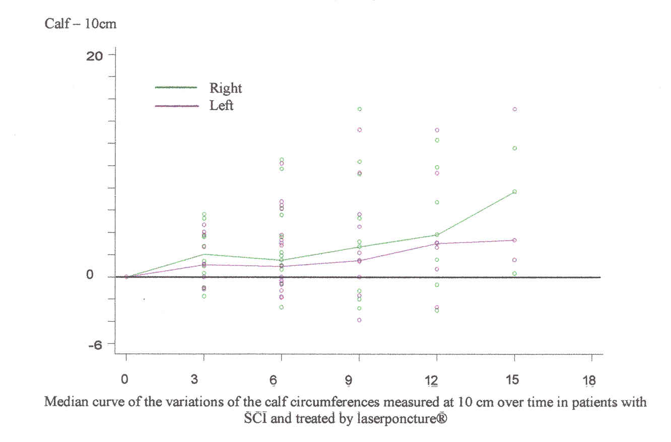
Descriptive statistical analyses were carried out in a blinded fashion. The measurements, expressed in percentage for each individual, were recorded over time. The system of reference “0” corresponds to the first measurement. The first variations, which appear at three months on the graph, correspond to the average of the variations observed between one and three months for each patient. The mechanism of calculation is the same for 6, 12 and 18 months.

The curve represents the median of these variations for all the individuals.

RESULTS:

In the graphs (below), the curves, representing the median of the variations of the thighs and calves measured at 10 cm and 15 cm, are coherent compared to each other.

The analysis shows that the right and left limbs do not change at the same time and at the same speed, but tend to balance each other.

1. The muscle volume of a limb can increase quicker than the other. It can be noticed that after the curves met, the tendency can be reversed and the limb, which was behind, can then increase quicker.

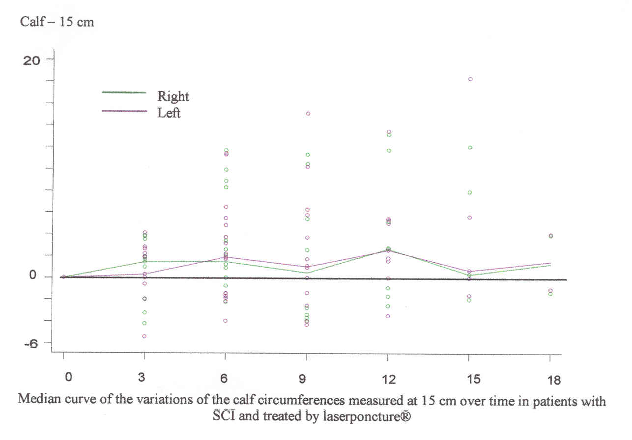
2. The two curves meet in the three graphs at 6, 12 and 18 months. This phenomenon begins towards the fifth month of treatment with the calves measured at 15 cm, i.e. the remotest measurement from the navel; then it evolves towards proximity with the calves measured at 10 cm towards the sixth month; and finally, the thighs towards the seventh month of laserponcture® therapy. Then, the curves of the lower limbs meet with a periodicity of six months on the three graphs.

The circumference of the calf measured at 15 cm evolves at its own pace of three months, which is faster than the calf and thighs measured at 10 cm. However, this variation alternates between rises and falls, and somewhat a global stability in measurements.

Whereas the evolution of the median value shows a rise in the curves, and thus a global rise of the volumes of the muscles, a certain number of points are below the system of reference “0” (first measure taken before any treatment). The points, situated below the system of reference, correspond to a reduction of the thighs and calves circumferences.

The maximal peak of reduction for calves and thighs measured at 10 cm is during the ninth month, if we compare to day 0. After twelve months, no measurement is below the system of reference. Thus, from the twelfth month onwards, the curve representing the median value of the circumferences variations of the calves and thighs measured at 10 cm speeds up.

DISCUSSION:

It appears that the right and left lower limbs in patients with SCI treated by laserponcture® have a progressive periodicity of six months, and tend to balance each other. Six and twelve months correspond to a peak for the measurement of the calf at 15 cm.

Between the sixth and ninth month, the body pauses in its recoveries. This stage is concomitant to the maximal negative data. The following period of three months shows a rise in the measurements. This stage, where there is a variation of circumferences in the calves and thighs measured at 10 cm, appears when the circumference of calves at 15 cm reduces.

Another significant stage in the evolution of measurements is between the twelfth and fifteenth month. Despite the low number of patients (7 represented for 12 months (31%), and 5 represented for 15 months (23%)), it seems that a speeding-up of the volumes of thighs and calves measured at 10 cm appear. It is concomitant with a reduction of the calf measured at 15 cm.

These progressive phenomena seem to have been triggered by variations of the most distal measurement, i.e. variation of the calf measured at 15 cm.

The latency period observed between the sixth and ninth month also corresponds to the maximal negative values. This stage could be a necessary time for the body to integrate the progress observed in the SCI patient morphology. Indeed, the negative values could express the progressive elimination of oedemas of stasis and thus mean an improvement of venous and lymphatic circulation with a diminution of the peripheral vascular resistances, which are two significant phenomena for the negative physiopathological evolution of a SCI individual (6, 8).

The improvement of vascular circulation seems to be the primus mouvens of any functional recovery and allows the muscles to increase their volumes.

CONCLUSION & PERSPECTIVES:

This study highlights the effects of laserponcture® therapy on the morphology of the lower limbs skeletal muscles in 22 patients with SCI regardless of their level of injury. The increase of the circumferences would mean a reversibility of muscle atrophy and return of some functionality.

These changes have occurred without the use of any electrical machine substituting for the motor control function, which is likely to damage the neuromuscular junctions. In the case of laserponcture®, a new start of a physiological process would begin with the lower limbs distality.

The progressive process is coherent, regardless of the points chosen in the patient’s cartography, the number and frequency of treatments, and the time since injury.

In the case of a scientific protocol, this technique can be performed by another therapist using the pre-established cartographies in order to show the efficiency of the therapy and its repeatability by a different team in a different context and with different patients.

Moreover, after these clinical observations, it would be interesting to look into details, such as functional MRI and histological assessment, and the transformation of paralyzed skeletal muscles because of SCI.

Finally, it is believed that laserponcture® therapy, applied as soon as possible after a SCI, will speed up the recovery process by avoiding the stage of the vascular modifications and shift of type I to type II muscle fiber, which could be a cause of spasticity.

GRAPHS:

1) Median curve of the variations of the thigh circumference measured over time in patients with SCI and treated by laserpuncture.

2) Median curve of the variations of the calf circumference measured at 10 cm over time in patients with SCI and treated by laserpuncture.

3) Median curve of the variations of the calf circumferences measure at 15 cm over time in patients with SCI and treated by laserpuncture.

*Authors:

Albert Bohbot, state-registered podiatrist, director and founder, Laboratoire de recherche sur le laserponcture®, independent researcher in neurosciences. Address: Château Gaillard – 33 route du canal – 18140 La Chapelle Montlinard – France – Phone and fax (work): +33 2 48 79 43 61 – Phone (home): +33 2 48 79 47 09 – Email: albert@laserponcture.net. Practitioner of laserponcture®.

Cécile Jame-Collet, MD, Traditional Chinese Medicine, Faculté de Médecine de Paris Nord. Address: Chérault – 58270 Saint-Benin-d’Azy – France – Phone: +33 3 86 58 45 18 – Email: cecile.jcollet@wanadoo.fr. Observer in charge of taking measurements.

ACKNOWLEDGEMENTS:

The assistive efforts of the following individuals are acknowledged: Dr. Laurance Johnston for critical comments, Edwige Nault for the translation, and Audur Gudjonsdottir for her commented reading.

BIBLIOGRAPHY:

1. Castro MJ, Apple DF Jr, Rogers S, Dudley GA. Influence of complete spinal cord injury on skeletal muscle mechanics within the first 6 months of injury. Eur J Appl Physiol 2000; 81 (1-2): 128-31.

2. Hopman MTE, Groothuis JT, Flendrie M, Gerrits KHL, Houtman S. Increased vascular resistance in paralyzed legs after spinal cord injury is reversible by training. J Appl Physiol 2002; 93 (6): 1966-72.

3. Shamir MH, Rochkind S, Sandbank J, et al. Double-blind randomized study evaluating regeneration of the rat transected sciatic nerve after suturing and postoperative low-power laser treatment. J Reconstr Microsurg 2001 Feb; 17 (2): 133-7.

4. Scelsi R. Skeletal muscle pathology after spinal cord injury: our 20 year experience and results on skeletal muscle changes in paraplegics, related to functional rehabilitation. Basc Appl Myol 2001; 11 (2): 75-85.

5. Butler JE, Thomas CK. Effects of sustained stimulation on the excitability of motoneurons innervating paralyzed and control muscles. J Appl Physiol 2003; 94 (2): 567-75.

6. Hesse S, Malezic M, Lücke D, Mauritz KH. Value of functional electrostimulation in paraplegics patients, Nervenarzt 1998 – 69: 300-305, Springer Verlag 1998.

7. Young W. Electrical Stimm? Dr Young. CareCure Community [forum online] 2003 May 5. Available from: URL: http://carecure.atinfopop.com/4/OpenTopic?a=tpc&s=4754088921&f=38540

88921&m=9324006872.

8. Bohbot A. Laserponcture®: an alternative of treatment for spinal cord injuries. Proceedings of Human Spinal Cord Injury: New and Emerging Approaches to Treatment; 2001 May 31-June 2; Reykjavik, Iceland. Available from: URL: http://perso.wanadoo.fr/laserponcture/english/congoms/bohbotus.htm

9. Bohbot A. Laserponcture®: an alternative treatment for spinal cord injuries. Proceedings of the 4th International Symposium on Experimental Spinal Cord Repair and Regeneration; 2002 March 25-27; Brescia, Italy. Available from: URL: http://perso.wanadoo.fr/laserponcture/english/symposium.htm

10. Jame-Collet C. Du dermatome au pibu, le lien: le laserponcture. Application clinique au travers de la para et de la tétraplégie [dissertation]. DUMETRAC. Univ. de Médecine Paris-Nord; 2001.

11. Johnston L. Laserponcture: the French connection. Paraplegia News 2000 Sep; 54 (9): 18-19.

12. Bohbot A. Laserponcture® theory and philosophy. Available from: http://www.healingtherapies.info/laserponcture-theory.htm

13. Johnston L. Conference report, human spinal cord injury: new and emerging approaches to treatment. Spinal Cord 2001 Nov; 39 (11): 609-613.

14. Johnston L. A matter of WHO. Paraplegia News 2001 Sep; 55 (9) 38-41.

15. Naeser MA and Deuel S.K. Review of second congress, world association for laser therapy meeting (WALT); 1998 Sep 2-5; Kansas City, MO, USA. J Altern Complement Med 1999 Apr; 5 (2): 177-80.

16. Lujer EJ, Rochkind S, Wollman Y, Kogan G and Dekel S (1998). Effect of low-power laser irradiation on the mechanical properties of bone fracture healing in rats. Lasers Surg Med 1998; 22 (2):97-102.

17. Rochkind S, Nissan M, Alon M, Shamir M and Salame K. Effects of laser irradiation on the spinal cord for the regeneration of crushed peripheral nerve in rats. Lasers Surg Med 2001; 28 (3): 216-9.

18. Rochkind S, Shahar A, Amon M and Nevo Z. Transplantation of embryonal spinal cord nerve cells cultured on biodegradable microcarriers followed by low laser irradiation for the treatment of traumatic paraplegia in rats. Neurol Res 2002; 24 (4): 355-60.

19. Wollman Y, Rochkind S, Simantov R. Low power laser irradiation enhances migration and neurite sprouting of cultured rat embryonal brain cells. Neurological Res 1996; 18: 467-70.

20. Wollman Y and Rochkind S. In vitro cellular processes sprouting in cortex microexplants of adult rat brains induced by low power laser irradiation. Neuro Res 1998 Jul; 20 (5): 470-2.

21. Naeser MA, Hahn K-A K, Lieberman BE, Branco KF. Carpal tunnel syndrome pain treated with low-level laser and microamperes transcutaneous electric nerve stimulation: a controlled study. Arch Phys Med Rehab 2002 Jul; 83 (7): 978-88.

22. Branco K, Naeser MA. Carpal tunnel syndrome: clinical outcome after low-level laser acupuncture, microamps transcutaneous electrical nerve stimulation, and other alternative therapies – an open protocol study. J Altern Complement Med 1999; 5 (1): 5-26.

23. Rochkind S, Ouaknine GE. New trend in neuroscience: low-power laser effect on peripheral and central nervous system. Neurol Res 1992; 14 (1): 2-11.

24. Rochkind S, Barr-Nea L, Razon N, Bartal A, Schwartz M. Stimulatory effect of He-Ne low dose laser on injured sciatic nerves of rats. Neurosurgery 1987 Jun; 20 (6): 843-7.

25. Rochkind S, Nissan M, Lubart R, et al.. The in-vivo nerve response to direct low-energy laser irradiation. Acta Neurochir 1988; 94 (1-2): 74-7.

26. Rochkind S, Nissan M, Barr-Nea L, Razon N, Schwartz M, Bartal A. response of peripheral nerve to He-Ne laser: experimental studies. Lasers Surg Med 1987; 7(5): 441-3.

27. Rochkind S, Volger I, Barr-Nea L. Spinal cord response to laser treatment of injured peripheral nerve. Spine 1990; 15 (1): 6-10.

28. Friedman H, Lubart R, Laulicht I, Rochkind S. A possible explanation of laser-induced stimulation and damage of cell cultures. J Photochem Photobiol B 1991 Oct; 11 (1): 87-91.

29. Anders J. from the Uniformed Services University of the Health Sciences, Department of Anatomy, Physiology and Genetics. Low Power Irradiation: Spinal Cord Injury. Available from: http://www.usuhs.mil/nes/Anders3.htm.

30. Nguyen Van Nghi, Mai Van Dong, collab. with J. Nguyen Viêt Bao. Hoang ti nei king so ouenn, Tomes I, II, III. Marseille: 1975.

31. Bossy J. Acupuncture, moxibustion, analgésie acupunctural; Synthèse des travaux des symposia de Pékin, 15 juin 1979. Paris: Ed. Doins; 1980.

32. Chusid JG. Manuel d’anatomie et de physiologie neurologiques. Paris: Ed. Masson; 1982.

33. Dittuno JF, et al. The International Standards Booklet for Neurological and Functional Classification of Spinal Cord Injury. Paraplegia 1994; 32: 70-80.