ABSTRACT:
Objective: To promote
additional functional recovery in patients with traumatic, incomplete
spinal cord injury (SCI).
Methods: An
intradural-lysis and peripheral-nerve implantation microsurgical procedure
was performed on 35 patients with obsolete or chronic (i.e., non-acute)
incomplete SCI. After the endorachis was opened, fibrous bands adhering to
the spinal cord from the arachnoid, pia mater spinalis, ligamenta
denticulatum, and initiating part of the nerve-root were removed. The
injury site was opened by three to six 0.1-0.2-mm-deep incisions that were
slightly longer than the abnormality area. The spinal cord cyst was
opened, and its liquid sucked off. Harvested autogenous, sural-nerve
segments were stripped of their spineurium and perineurium - making them
resemble cauda equine tissue - and implanted into the incisions. Finally,
the spinal cord was sutured with pia mater spinalis, and the endorachis
was sutured over with a sacrospinal muscular pedicle flap.
Results: Patients were
postoperatively followed for a period ranging from 1 to 6 (average 2.5)
years. In 32 of 35 cases, sensory and motor function increased at least
one grade using the Frankel SCI classification scale; the other three
cases recovered only one sensation grade. Of the 32 cases, main upper-leg
muscle strength improved one grade in 23 cases and two grades in nine
cases. All 32 recovered some ambulatory ability.
Conclusion:
Removing the endorhachis adhesions, cutting into the cicatricial spinal
cord, and implanting autogenous peripheral-nerve segments enhanced
functional recovery in individuals who have obsolete paralysis caused by
incomplete SCI.
INTRODUCTION:
Obsolete or chronic paralysis caused by traumatic SCI
remains a challenging problem. Although many post-injury changes in the
spinal cord can be readily observed with imaging technology, such as MRI,
an individual with SCI may, nevertheless, have a near-normal MRI.7
Indeed, apart from compression and instability factors, patients with
similar MRI changes may have much difference in functional recovery.
Through anatomical and surgical observations, we believe that the main
factors in these differences are adhesions within the endorachis,
resulting, for example, from fibrous strip traction and the traumatic scar
and cyst’s overall physical characteristics, including
degeneration-related mollescence. Since 1994, we have treated 35 patients,
whose functional recovery, we believe, was inhibited by such factors, by
microsurgical intradural-lysis and peripheral-nerve implantation
procedures. Results indicate that the procedures promoted additional
functional recovery.
CLINICAL MATERIAL & METHODS:
Patient Population:
The study group of 35 patients with obsolete or
chronic paraplegia (i.e., non-acute) included 30 males and 5 females.
Their age ranged from 16 to 42 (average 31) years. Ten, nineteen, and six
patients were injured in the thoracic T7-T9 region,
T10-T12 region, and lumbar L1-L2
region, respectively. Seven had been injured from falls from high
places, five from heavy objects crashing down on them, three from gunshot,
two from knife, and 18 from traffic accidents. The time interval from
injury to the surgical intervention ranged from 6 to 26 (average 13)
months.
Before the subject surgical intervention (i.e., in
the acute stage), decompression and bone-grafting and internal fixation
procedures (24 by pedicle screw; 11 by Z plate) had been carried out in
all patients. In sixteen, the internal fixation devices had been removed
before the microsurgical intervention. In addition, 11 patients were
treated with hyperbaric oxygen. Although imaging indicated no compression
or instability, three months after decompression and fixation functional
recovery had ceased in all patients and remained so until the
intradural-lysis and nerve-implantation procedure.
According to the Frankel impairment classification
scale,4 31 patients were initially level B, and 4 were level C.
(In this scale, A represents a complete injury and E normal function; B
through D represent incomplete injuries).
Operative Technique:
Patients were placed in a lateral position and
subjected to general anesthesia. After making a midline incision, the
endorhachis was exposed and opened with the assistance of a 4-6X forehead
microscope. The arachnoid, pia mater spinalis, initiating nerve-root
component, and the space between the front and back branch were observed
carefully for bands, strips, scars, or adhesions, and for any area
affected by ligamenta-denticulatum dragging. Because such elements were
small, careful, repeated observations were necessary to ensure therapeutic
effectiveness.
The initial part of the nerve root routinely adhered
to the spinal cord, with a strip between the front and back nerve-root
branch that dragged or pinched the cord. In addition, the adhesion of the
arachnoid and the pulling of the ligamenta denticulatum transformed the
spinal cord. For example, the pia mater spinalis became thicker and, as a
result, adhered and compressed the cord. Adhesions between the spinal cord
and the arachnoid, pia mater spinalis, ligamenta denticulatum, nerve root,
as well as the peripheral fibrous strip were completely relieved.
After lysis, the injured cord area was opened by
three to six 0.1-0.2-mm-deep incisions that were slightly longer than the
injury area. If the cyst was one-cm2 or bigger as indicated by
the pre-surgical MRI or could be clearly observed through its
dark-colored, undulant, and thin-walled nature, it was punctured and
incisioned, and its fluid sucked off.
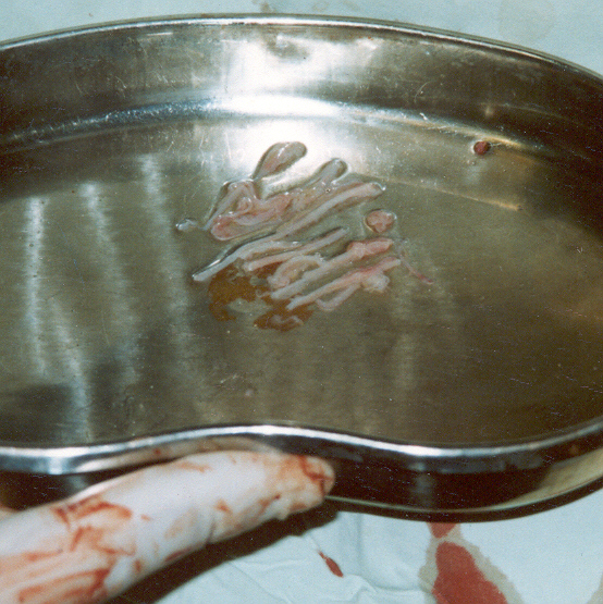
Autogenous sural-nerve segments were harvested
corresponding to the length of the area of abnormality. After the segments
were micros urgically
denuded of their spineurium and perineurium - making them resemble cauda
equine tissue, they were implanted into the aforementioned incisions and
cyst cavity. Finally, the opened spinal dural was sutured with the pedicle
muscular flap and the incision covered with sacrospinal muscle.
urgically
denuded of their spineurium and perineurium - making them resemble cauda
equine tissue, they were implanted into the aforementioned incisions and
cyst cavity. Finally, the opened spinal dural was sutured with the pedicle
muscular flap and the incision covered with sacrospinal muscle.
RESULTS:
Patients were postoperatively followed for a period
ranging from 1 to 6 (average 2.5) years. In 32 of 35 cases, sensory and
motor function increased at least one grade using the Frankel SCI
classification scale; the other three cases recovered only one sensation
grade. Of the 32 cases, main upper-leg muscle strength improved one grade
in 23 cases (21 from Frankel B to C, two from C to D) and two grades in
nine cases (7 from Frankel B to D and two from C to E). All 32 patients
recovered some ambulatory ability. The patients who improved two grades
could walk without crutches, and, in the case of the two that improved
from Frankel C to E, near-normal function was acquired. In 21 cases, bowel
and bladder function clearly improved. Of the six cases who suffered
serious nerve-root pain before the operation, pain was completely
alleviated in two, slight pain remained in two, and little improvement
noted in two.
DISCUSSION:
Rationale for Intradural Lysis: Normally, pia
mater spinalis is soft and rich in blood vessels. It clings to the spinal
cord’s surface and goes deep into the fissurae mediana anterior at the
cord’s front side. Together with nerve roots, it goes through subarachnoid
space and connects to the endorachis. On the side surface, there are two
rows of triangle ligaments called ligamenta denticulatum, which present
two lamina-like layers, starting from the foramina magnum and reaching to
the terminal cone at the first lumbar vertebral level.
On the ligamenta denticulatum’s external surface, 19
to 21 dentations extend from the pia mater spinalis in a sawtooth fashion.
Their tips push the arachnoid to the outside, and attach to the endorachis’
inner surface between the upper and lower nerve root, fixing the spinal
cord. Hence, the pia mater spinalis, arachnoid, two neighboring nerve
roots, ligamenta denticulatum between two nerve roots, and the endorachis
constitute a relatively separate unit.2
When the spinal cord is injured, dural sac blood will
adhere to these tissues, the fibrous scar, and the cord.7
Various spinal-cord restricting adhesions were present at the traumatic
area in all cases. Spinal-cord deformity caused by harmful ligamenta
denticulatum stress traction could also be seen.
The anterior and posterior radicular arteries supply
the cord’s blood, and play a compensatory role when the anterior and
posterior arteriae mediana are injured. However, when the anterior and
posterior radicular arteries as well as the anterior and posterior
arteriae mediana are injured or pressed at the same time, the blood supply
will be severely compromised, which, in turn, aggravates the injury.
Because the injury area overlaps with surrounding
structures and because the scar is thin and slender, function-inhibiting
adhesions cannot be clearly seen by CT or sagittal MRI imaging, and the
coronary MRI reveals little. Hence, pathological changes cannot be
documented by imaging techniques.7 These changes cannot be
relived by routine fracture reduction, decompression, and fixation
procedures because they are routinely undertaken outside the endorachis
(Fig 4).
Rationale for Peripheral Nerve Implantation: Because under routine
circumstances, an injured neuron cannot be replaced and a damaged axon
cannot readily reproduce,6 it is difficult for appropriate
anatomical and functional connections to be reestablished. Because some
intact neurons remain in an incomplete injury, a transplant will bridge
them and injured axons, enhancing axonal regeneration potential, and, in
turn, spinal-cord functional recovery.8
The
muscles affected by the necrosis of the anterior gray column cells of the
1st-2nd segments may not be completely paralyzed. However, in a pyramidal
tract injury, all muscles controlled by the injured and lower-segment
spinal cord are paralyzed. As such, it is important for a potential
treatment to accelerate the regeneration of the long-conductive bind and
to create regeneration-enhancing conditions for the spinal cord.
Ideally, a graft should not only have bridging capabilities but also be
able to provide the appropriate extracellular matrix components and
cellular trophic factors conducive to neuronal regeneration.
We
believe that autogenous peripheral-nerve segments are good grafting
candidates. First, after an incomplete injury, surviving neurons are
isolated from each other in islands of non-functional cell mass separated
by intervening dead neuronal or micro-scar tissue. If connections can be
reestablished between surviving neurons, some conduction through the
injury site will be restored. As such, the purpose of our transplantation
procedure is to enhance axonal regeneration through the graft’s guiding,
bridging, and matrix-supporting characteristics. Such axonal regeneration
will help connect the cell-mass islands, forming synaptic junctions
between the cord’s contused ends and, in turn, enhancing conduction and
functional recovery.
Second, our microsurgically prepared grafts provide the extracellular
foundation and growth factors to trigger the axonal regeneration that
forms the basis for regeneration of the long conductive strip. Studies
suggest that the cord’s long-conductive strip neurons can penetrate the
graft-host interface and form extensive connections that enhance the
cord’s functional recovery.
In
contrast to other grafting procedures, the spineurium and perineurium of
our grafts have been removed, which allows the spinal cord to directly
connect and interact with nerve fibers, glial cells, and other beneficial
factors that exert guiding effects for regenerating nerve cells. 5,3
Third,
our microsurgically prepared grafts contain important cells, such as
Schwann cells and fibroblasts, and a variety of important trophic factors.
Because these peripheral-nervous tissue elements are not readily flushed
away from the injury area by cerebrospinal fluid, a sustained effect is
observed.
Fourth, we believe that our procedures are superior to olfactory or fetal
nervous tissue transplantation, or the exogenous or endogenous application
of nerve-growth factors. By providing an excellent environment for
neuronal regeneration, our grafts promote spinal-cord functional recovery.
6 Other advantages include immunological acceptance, ready
integration of the graft into the host tissue, no need for highly
specialized graft-preparation methodology, and a relatively
straightforward, clinically generalized surgery.
Indications for Operation: As mentioned
earlier, surgical indications were incomplete paraplegia that
showed some functional recovery soon after injury but reached a plateau
three months post-injury that continued for an additional three months.
Provided that these cases showed no evidence of bone compression, canales
spinalis stenosis, or spinal unsteadiness, the injured cord should be
surgically examined, and scar tissue completely loosened. As a result, the
spinal cord compression is alleviated and blood flow improved.
The basis for the time of operation was that after
bone compression and instability was alleviated, the incompletely injured
spinal cord had lived through microcirculation disturbance and edema, and
nerve function reached its first recovery peak. At the same time, the scar
tissue began to grow and reached its peak after three months. If the scar
compressed the spinal cord, recovery would cease or even decline. 1,3
Because three months later the scar began to soften
and partly be absorbed, the compression gradually vanished, and the second
recovery peak appeared. If the cord’s functional recovery completely
ceased at this time, it was concluded that the scar-tissue compression
could not be relieved by the body itself; hence, the intradural-lysis
procedure should be performed.
For cases who showed no evident bone compression,
canales spinalis stenosis and spine unsteadiness in MRI and CT images, yet
whose spinal functional recovery had ceased, the compression in the dural
sac is likely the main cause. The diminutive scars and fibrous strips are
difficult to be observed with imaging techniques. Because they cling to
the cord and there is no buffering action of the cerebrospinal fluid and
fat as there is outside the endorachis, the influence of these tissues is
more direct and serious.
It is always observed that the scar tissue in the
dural sac forms laterigrade or tilted strips, compressing the cord. The
spinal pulse is observed at the proximal site and disappears at the distal
site; after getting rid of the strips, the pulse reappears. Although the
intradural-micro-lysis procedure is efficacious, it is neglected in the
routine decompression operation, which may help explain the operation’s
poor results in some cases. Because our procedures are performed in the
dural sac, the surgeon must have rich micro-neurosurgery experience to
prevent further functional loss.
CONCLUSION
The main objective of most decompression operations
is to eliminate outside compression of the dural sac and stabilize the
spine. Intradural scar and adhesion influences on spinal cord functional
recovery are often ignored. By completely loosening such scars and
adhesions, our microsurgical procedures eliminate these
function-inhibiting influences. As a result, significant improvement was
noted in all treated patients.
TOP