I was invited to speak at the 5th
International Symposium on Experimental Spinal Cord Repair and
Regeneration about various therapies discussed in my PN “Healing
Options” series. Held on March 27-29 in Brescia, Italy, the Symposium was
hosted by University of Brescia’s Dr. Giorgio Brunelli.
Believing
 that
the development of new therapies for spinal cord injury (SCI) requires
global and multidisciplinary cooperation, organizers invited scientists
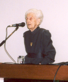
with diverse viewpoints from throughout the world. As in the previous
symposium, a highlight was the participation of Dr. Rita Levi-Montalcini,
the 1986 Nobel Laureate in Medicine for discovering nerve growth factor
over 50 years ago. Following are summaries of a few presentations:
that
the development of new therapies for spinal cord injury (SCI) requires
global and multidisciplinary cooperation, organizers invited scientists
with diverse viewpoints from throughout the world. As in the previous
symposium, a highlight was the participation of Dr. Rita Levi-Montalcini,
the 1986 Nobel Laureate in Medicine for discovering nerve growth factor
over 50 years ago. Following are summaries of a few presentations:
Neuromonitoring:
A keynote lecture was given by Dr. Milan Dimitrijevic
(Texas), an internationally recognized neurophysiologist whose
investigations on spinal-cord conduction have greatly contributed to our
understanding of SCI. He noted that the spinal cord is much more
physiologically complex than originally thought; however, rather than
making functional repair more challenging, this complexity generates
opportunities for new rehabilitation strategies.
Dimitrijevic discussed how various interventions
after acute SCI - such as surgical stabilization and reconstructions -
could be monitored by recording conduction above and below the injury
site. By using such neuromonitoring, a surgeon can have valuable
information on where spinal-cord integrity is being preserved.
Through knowledge gained from such monitoring, it may
be possible to initiate physiological-like electrical stimulation of the
non-injured conducting axons to prevent effects of disuse and secondary
lesions.
In chronic injuries, neuromonitoring could assess the
efficacy of various function-restoring interventions. For example, the
neuromonitoring of bridging tissue transplants could provide information
about whether the transplants are filling the gap, integrating with
surviving tissue, decreasing scar tissue, reducing cavity formation,
staying where placed, and increasing migration of blood vessels or cells
that facilitate axonal regeneration.
Rerouted Nerves:
Brunelli, the symposium organizer, reviewed his
research on surgically rerouting human peripheral nerves (i.e., those
outside of the spinal cord and brain) around the injury site to
reestablish functional neuronal connections. For example, he has restored
some function by redirecting the wrist’s ulnar nerve and connecting it to
nerves that control leg functioning below the injury site. After this
procedure, a patient with a complete spinal-cord transection could stand
up and walk short distances.
In another procedure carried out in a woman with a
complete thoracic transection, the peroneal nerve (to the leg) was used as
a bridge directly from the spinal cord above the injury site to the nerves
of the gluteus and quadriceps muscles. After two years, she was able to
walk 30-40 meters with a walker.
Because the second procedure represents a direct
peripheral-nerve-to-spinal-cord connection, it challenged traditional
beliefs on how neurons control muscle function. Specifically, in the
previous symposium, Levi-Montalcini was troubled by the implications of
Brunelli’s work because upper motor neurons (nerves within the spinal
cord) and lower motor neurons (nerves that leave the cord to connect to
muscles) use different neurotransmitters (chemicals released from a neuron
ending that interacts with an adjacent neuron or muscle cell). Hence,
theoretically, the muscle should not be triggered due to neurotransmitter
incompatibility.
Because scientists pay special attention to the
suggestions of Nobel laureates, Brunelli and collaborators have since
shown that this procedure indeed restores function in rats in spite of
this putative incompatibility. Specifically, they have demonstrated that
the target muscles are genetically reprogrammed, producing receptors that
are responsive to the neurotransmitters released by the upper motor
neurons that have grown to the muscles through the peripheral nerve
bridge.
Radiation:
Dr. Nurit Kalderon (New York) has treated acutely
injured rats with radiation, which destroys nascent scar-tissue that
blocks neuronal regeneration. Although the spinal cord attempts to repair
itself soon after injury, the decay process takes over after the third
week. However, when x-rays were directed to the transected spinal cord
during the third week, the cord continued to repair. Once the wound was
healed, severed neurons grew across the site, restoring some function.
Because x-rays destroy obstructive cells, spinal-cord repair can continue.
Kalderon carried out additional studies in rats
injured by contusion, which more closely resembles most human injuries.
Before radiation, surgery was performed to reduce secondary damage caused
by swelling and fluid buildup. Starting 12 days postinjury, the lesion
was radiated daily for 10 days at a level proportionate to that clinically
used to remove human cancer. Analysis indicated significant tissue
repair.
Activity-Mediated Neurorehabilitation:
Dr. Humberto Cerrel Bazo (Italy) discussed the use of
activity–mediated training - such as functional electrical stimulation (FES)
- to maximize function after SCI. His talk, as well as others, referred to
the spinal cord’s “central pattern generator,” which can sustain
lower-limb repetitive movement, such as walking, independent of direct
brain control.
Cerrel Bazo noted that we are learning much more
about 1) the plasticity (adaptive mechanisms by which the nervous system
restores itself) of the sensorimotor nervous system above and below the
injury site (i.e., brain-spinal-cord-motor unit), and 2) the residual
activity of muscles, bladder, bowel, etc. amenable for enhanced function
through new circuitry or artificial means.
He reviewed how a sacral-sparing assessment procedure
(gauges anal sensation and control) within 30-days of injury is useful for
predicting future functional recovery.
Through FES training, Cerrel Bazo has shown
promising, plasticity-associated outcomes, which, in turn, suggests
intriguing possibilities about the integration of different systems above
and below the injury site. FES may enhance residual potential in people
with chronic SCI by generating a movement pattern useful for standing,
stepping, and cycling.
However, if residual activity remains dormant over
the long term, awareness to support activity may be lost. A properly
stimulated sensorimotor nervous system may generate activity useful for
the functional integration of different systems. In this sense, FES, the
awareness-learning process, and training mediated-activities may open
communication pathways between areas above and below the injury site.
Macrophage Therapy:
Drs. Eti Yole, Nachson Knoller, (Israel), and Sir
Jacques Brotchi (Belgium) summarized Proneuron Biotechnologies’ efforts to
use activated macrophages (a white blood cell) isolated from patient’s
blood to minimize neurological damage after acute SCI. Although healing
immune cells are scarce in the “immune-privileged” central nervous system,
Proneuron has circumvented this limitation by incubating the patient’s
macrophage-containing blood with skin tissue. The isolated macrophages are
then surgically implanted into the spinal cord within 14 days of injury.
By mediating protective immune responses, these activated macrophages
promote functional recovery.
Phase 1 clinical trials showed no adverse treatment
effects. Functional improvement was measured using the American Spinal
Injury Association (ASIA) assessment standards. Of the eight patients
treated (2 cervical and 8 thoracic injuries), three improved from ASIA A
to C (complete injury to partial motor and sensory recovery). Of the
remaining ASIA A patients, three showed improvements in sensory scores and
nerve conduction. Proneuron has initiated a much larger phase II clinical
trial that will recruit 61 patients at five treatment sites: Sheba
Hospital (Israel), Craig Hospital (Colo.), Mt. Sinai (N.Y.), Kessler
Rehabilitation (N.J.), and Shepherd Center (Ga.).
Conclusion:
As reflected by symposium presentations, the once
considered insurmountable barriers to restoring function after acute or
chronic SCI are gradually being eroded in many ways. More importantly,
over time, there has been a cumulatively huge shift in the attitude of the
inherently conservative scientific community. Namely, SCI is no longer
automatically a life sentence of paralysis without parole but a
neurological disorder amenable to function-restoring therapies. This shift
in the scientific collective consciousness by itself will greatly
accelerate the development of real-world therapies.
Adapted from article appearing in Paraplegia News (For subscriptions,
call 602-224-0500) or go to www.pn-magazine.com).
TOP